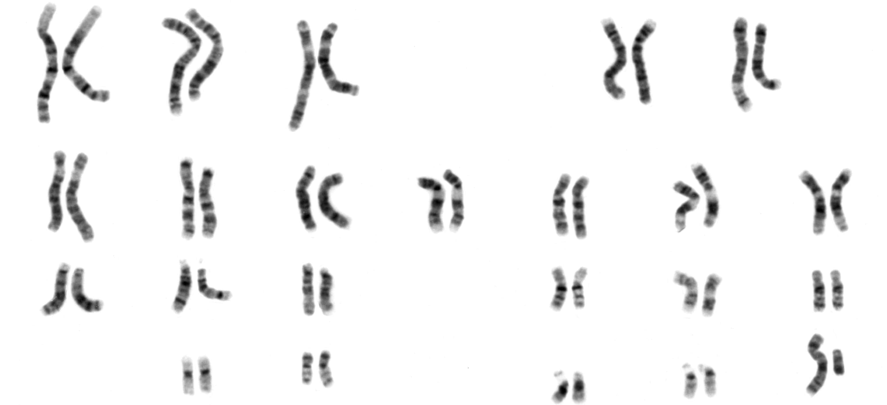
Karyotyping is the process of pairing and ordering all the chromosomes of an organism, thus providing a genome-wide snapshot of an individual’s chromosomes. Karyotypes are prepared using standardized staining procedures that reveal characteristic structural features for each chromosome. Clinical cytogeneticists analyze human karyotypes to detect gross genetic changes—anomalies involving several megabases or more of DNA. Karyotypes can reveal changes in chromosome number associated with aneuploid conditions, such as trisomy 21 (Down syndrome). Careful analysis of karyotypes can also reveal more subtle structural changes, such as chromosomal deletions, duplications, translocations, or inversions. In fact, as medical genetics becomes increasingly integrated with clinical medicine, karyotypes are becoming a source of diagnostic information for specific birth defects, genetic disorders, and even cancers.
Preparing Karyotypes from Mitotic Cells
Karyotypes are prepared from mitotic cells that have been arrested in the metaphase or prometaphase portion of the cell cycle, when chromosomes assume their most condensed conformations. A variety of tissue types can be used as a source of these cells. For cancer diagnoses, typical specimens include tumor biopsies or bone marrow samples. For other diagnoses, karyotypes are often generated from peripheral blood specimens or a skin biopsy. For prenatal diagnosis, amniotic fluid or chorionic villus specimens are used as the source of cells.
The process of generating a karyotype begins with the short-term culture of cells derived from a specimen. After a period of cell growth and multiplication, dividing cells are arrested in metaphase by addition of colchicine, which poisons the mitotic spindle. The cells are next treated with a hypotonic solution that causes their nuclei to swell and the cells to burst. The nuclei are then treated with a chemical fixative, dropped on a glass slide, and treated with various stains that reveal structural features of the chromosomes.
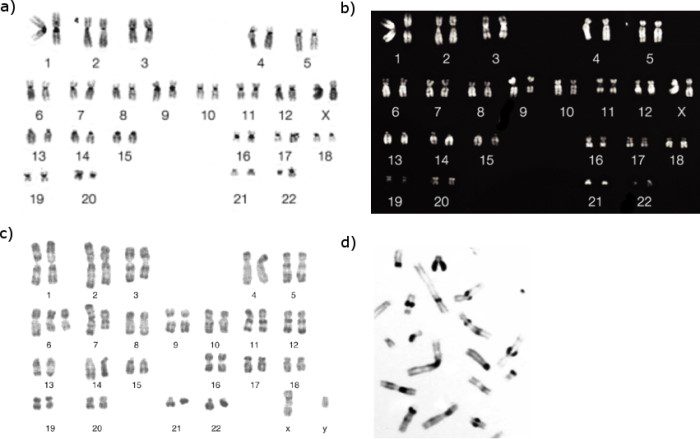
Chromosome banding revealed by different staining techniques.
Different chromosomal staining techniques reveal variations in chromosome structure. Cytogeneticists use these patterns to recognize the differences between chromosomes and enable them to link different disease phenotypes to chromosomal abnormalities. Giemsa banding (a), Q-banding (b), R-banding (c) and C-banding (d) are shown.